
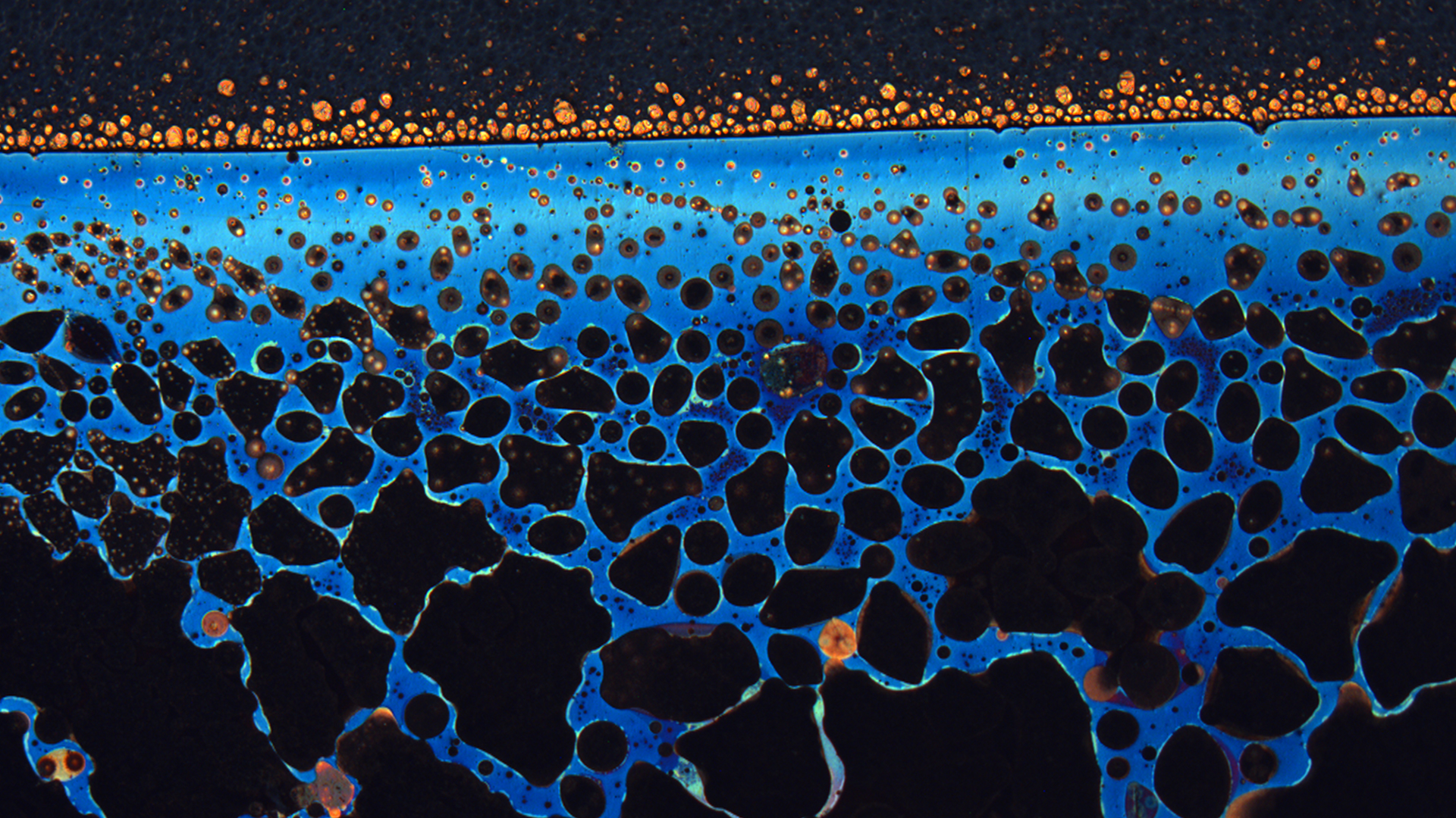
Copper beach
This image shows the degradation of an exposed film of copper in contrast with a thin protected layer that has not degraded—creating a picture resembling the blue waters and sandy shoreline of a beach. The small area at the top of the image has maintained its natural copper colour thanks to a protective coating. We investigated the ability of this biocompatible coating by laminating it over the surface of 100-ångstrom-thick evaporated copper on a glass substrate (the dark portion at the top of the image) and then exposing the sample to 100% relative humidity. This extreme level of humidity accelerated the corrosion of the exposed copper, forming this blue lace-like network, which preserved the copper’s conductivity. The laminated section of copper did not corrode as visibly, and this was reflected by better electrical conductivity, which shows that the biocompatible protective coating was successful. My research focuses on investigating the relationship between molecular structure and the physical properties of biocompatible films and next-generation materials. The aim is to create a library of eco-friendly protective materials to keep our planet safe and clean.

Photo by Antoine Durocher
Luming Fan
Patrizio Vena
Jeffrey M. Bergthorson
Marc Füri
Gilles Bourque
David May
Julien Sirois
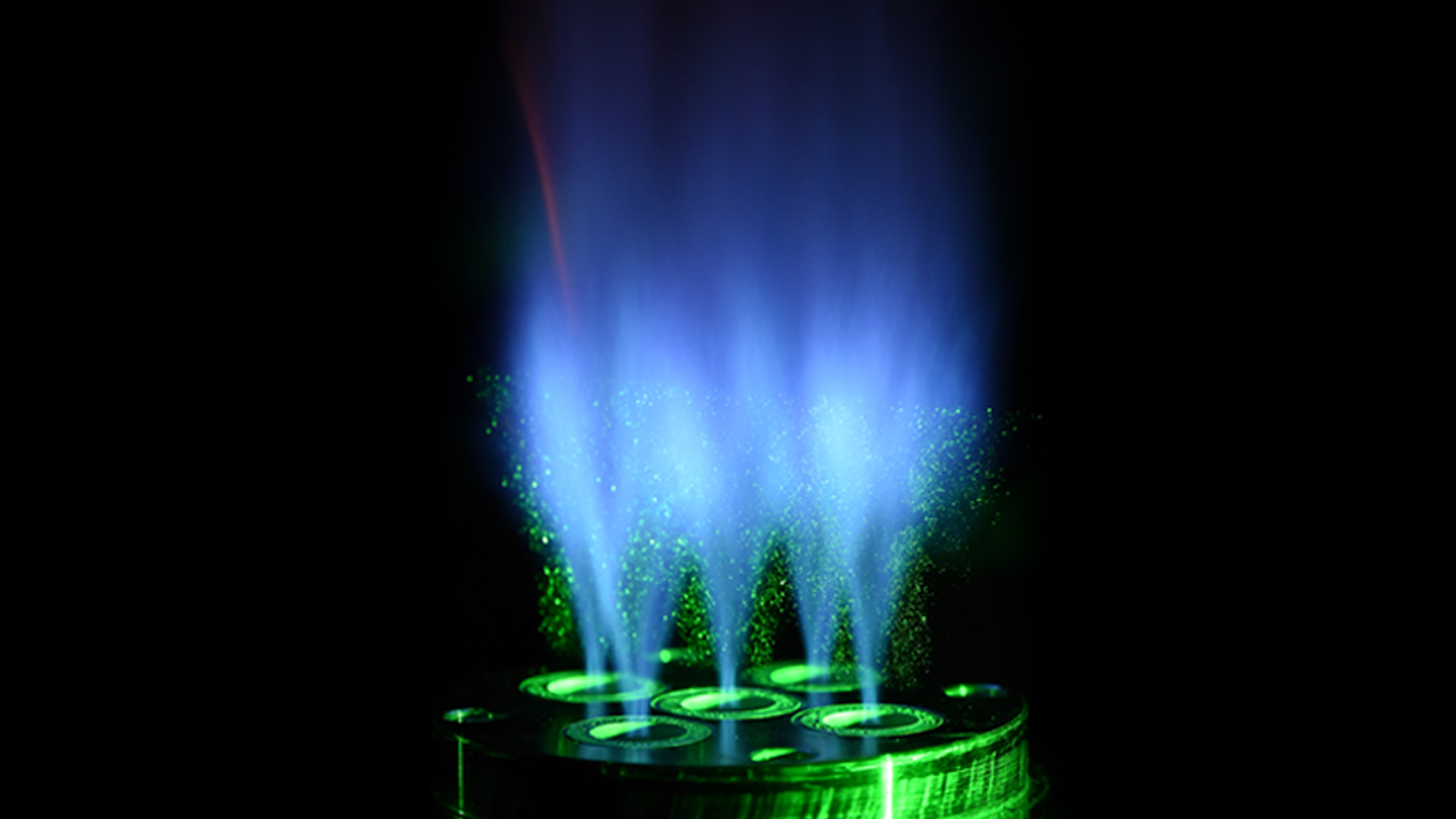
High-hydrogen content flames in 3D-printed micromix burners
Luming Fan
Patrizio Vena
Gilles Bourque
David May
Julien Sirois
Hydrogen alone burns without any carbon emissions, but it is intrinsically more unstable than current hydrocarbon fuels. Equipment can be damaged by using traditional combustion technology to burn high-hydrogen content fuels. This image shows a novel 3D-printed burner being used to stabilize high-hydrogen content flames into pure hydrogen. The burner relies on micromix technology—internal miniature fuel channels that are printed in the burner injector body to deliver fuel to localized sites. The five injectors are placed in a plus-sign shape to mimic the conditions of a hot gas turbine for the center flame. Then, we use non-intrusive laser diagnostics to investigate the combustion phenomena of these micromixed, high-hydrogen content flames and identify promising low-emission injector designs.

Nanoplastics: a major environmental issue
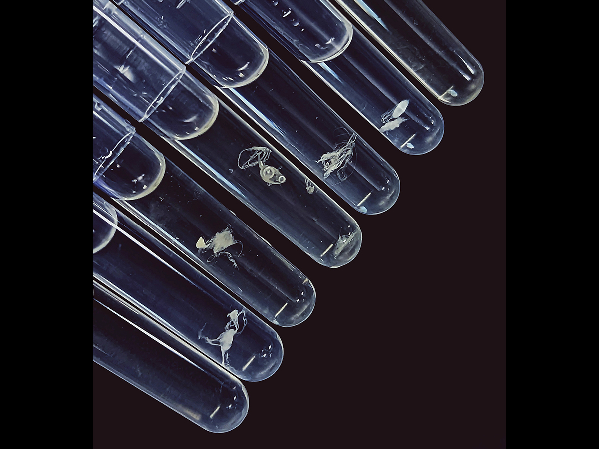
Nanoplastics (NPs) are found all over the world, and research that tackles this major environmental problem is urgently needed. This image shows NPs (yellow-green) in a water flea (Daphnia magna). We used confocal microscopy to locate the NPs with their intensity signal. D. magna are ideal for use with fluorescent-based microscopy techniques, because their bodies have blue autofluorescence. To achieve this image, we exposed D. magna neonates to 20 nm fluorescent polystyrene NPs (yellow-green) for 24 hours (acute toxicity). We can see the internalization of NPs in the gut, as well as some gut disruption (fragmentation) caused by NP uptake. NPs can also be seen in the antennae and claws, which D. magna use to navigate the water. The thoracic limbs are also stained with a yellow-green colour, indicating that NPs were retained as part of the filtration process.

The roots of education
Quebec is experiencing a remarkable boom in outdoor education. The benefits of this field have been widely documented, both in terms of contact with nature and on the cognitive, physical, psychological and social levels. However, new data collection tools are needed to assess learning. In December 2022, a team from the University of Sherbrooke spent a day observing first graders in Ms. Marie-Line's class at the Académie des Sacrés-Cœurs. The two children in the foreground are methodically marking out a gnome trail for a pre-Christmas math activity. One meter in a straight line maximum, then you branch off! That's the rule with gnomes...
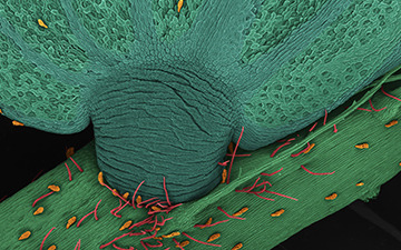
Plant reflex
The simple brush of an insect is enough to trigger an astonishing defensive reaction in the modest mimosa. Within a mere second, the leaflets, which make up the leaf, close one against the other. The extraordinary amplitude of this movement is provided by a leaf pad (dark green), the pulvinus. This structure works like a hydraulic robotic arm. A rapid transfer of internal fluids causes a portion of the pulvinus to deflate, shortening it, while, on the other hand, the sudden inflation lengthens its wall. A mechanism to be filed under biomimicry!

The veil of death
The shining star at the bottom of the image is quite young. It is about 5 million years old, while our Sun has been around for 5 billion years. However, WR 136 is about to take its last breath. With an initial mass estimated at 50 suns, it has already used up all its hydrogen, hence its initial collapse. As a result, it has ejected about half of its mass in the form of a gas bubble that is 30,000 times the size of our solar system. In blue, the particles moving towards us, and in red, those pulling away. WR 136, which is now burning its helium, is condemned to ultimately explode as a supernova or to end up as a black hole.

Spinning widow
After detecting vibrations in its web, this western black widow scurries to discover the source. In the Latrodectus Hesperus species shown, some females are more aggressive than others, and this is reflected in the architecture of their web. Thus, a spider quick to attack its prey will turn its web into an effective trap by weaving in numerous sticky threads. Conversely, a less aggressive female will weave a defensive web with threads firmly anchored to the ground, more likely to fend off aerial predators. This is a surprising discovery by the researcher and her team, who study animal personality.

18lets
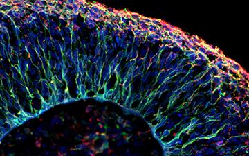
This image shows interneuron populations in a mouse’s cerebellum. Interneurons are a type of brain cell in the nervous system that send signals between themselves, and with both sensory and motor neurons in the brain. We used naturally occurring viruses which have a propensity to target and transfect brain cells to deliver fluorescent proteins to these cells. With images like this, we can study the function (i.e., what they do) and morphology (i.e., what they look like) of certain cells. In this image, the morphological profiles of inhibitory interneurons were labelled using black fluorescent proteins, and a subset of these interneurons have their nuclei labelled in red. We hope that this research will allow us to gain a deeper understanding of how the brain wires into functional circuitries and organisms during development.

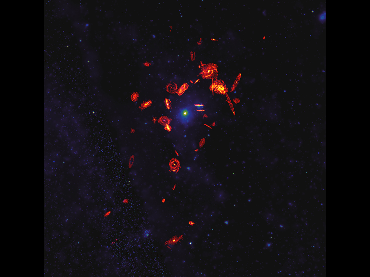
A cosmic whodunit in the Virgo Cluster
This image shows the million-degree plasma glow of the Virgo Cluster, a particularly extreme region of space. The star formation of galaxies within this cluster is being shut down; galaxies are being killed, and astronomers want to know why. Our research focuses on 49 galaxies that, along with thousands of others, are moving at millions of kilometres per hour through the cluster, living and dying by the violent physical processes of their environment. We use data to identify those processes and learn how they affect the life cycle of galaxies by observing the destruction of the vast reservoirs of dense gas from which stars are formed. In studying beautiful images like this, we find that external physical processes are capable of reaching far into the inner parts of galaxies to strip their star-forming gas, dramatically affecting how galaxies evolve as they fall into the cluster.

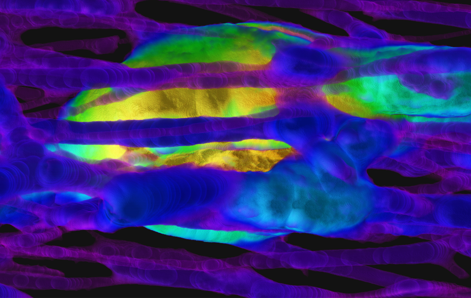
A living record
A biographer of our lifespans and biological archive, bone is a dynamic living tissue that constantly changes over individuals’ lives while recording life history information along the way. During the remodelling process, teams of bone cells can adapt based on adjustments to our diets, activity regimens and the substances we ingest (e.g., drugs). This high-resolution 3D render of bone was generated using Synchrotron Micro-Computed Tomography at Canada’s only synchrotron facility: the Canadian Light Source. This render visualizes the thickness of bone’s vascular canals (pictured in blue/purple) and provides a snapshot of the bone remodelling process (gold orb), during which bone cells demonstrate their remarkable ability to adapt to ever-changing physiological demands, leaving behind a preservable and readable record even centuries after death.

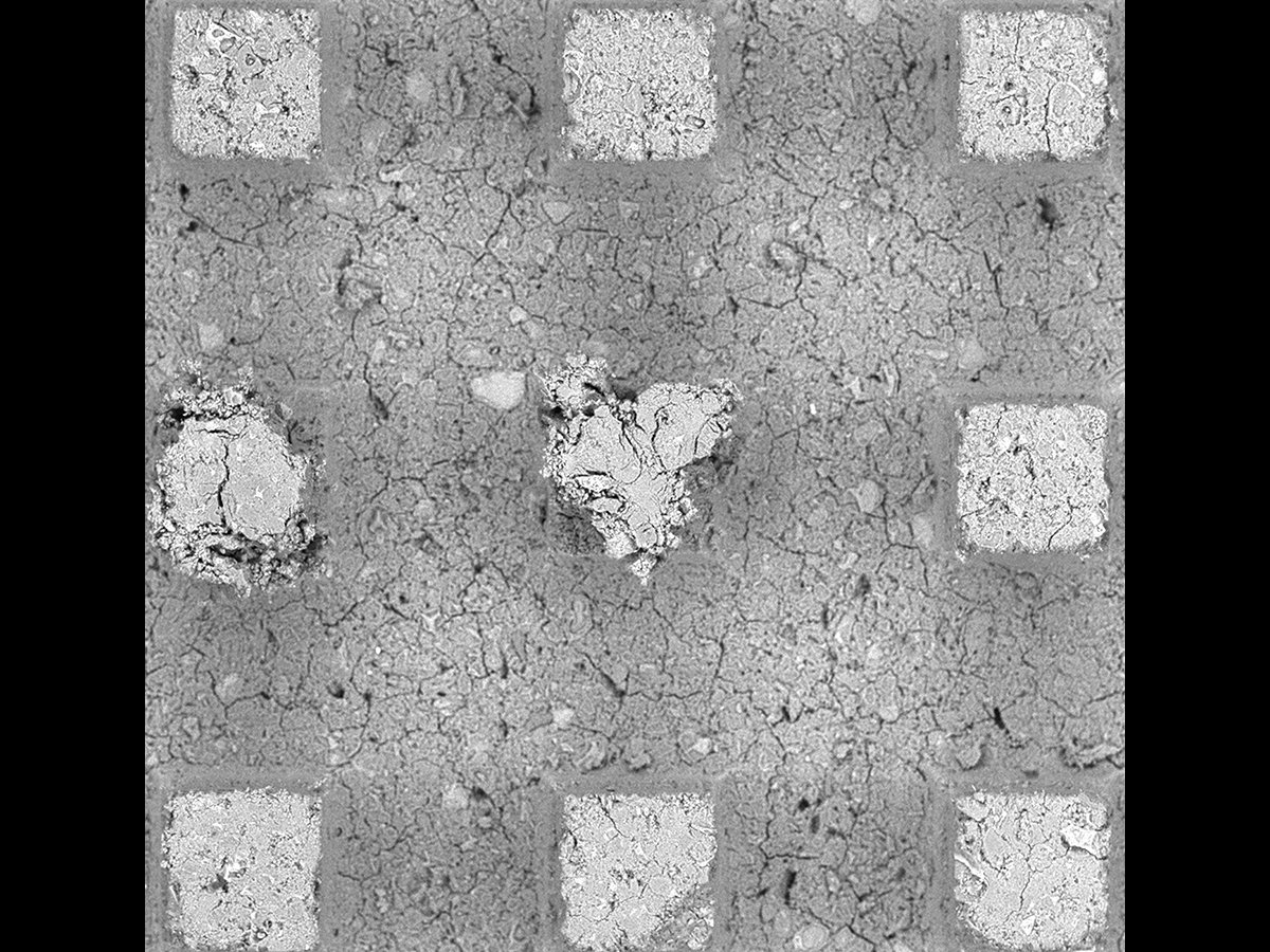
Cement paste micro heart
Luca Sorelli
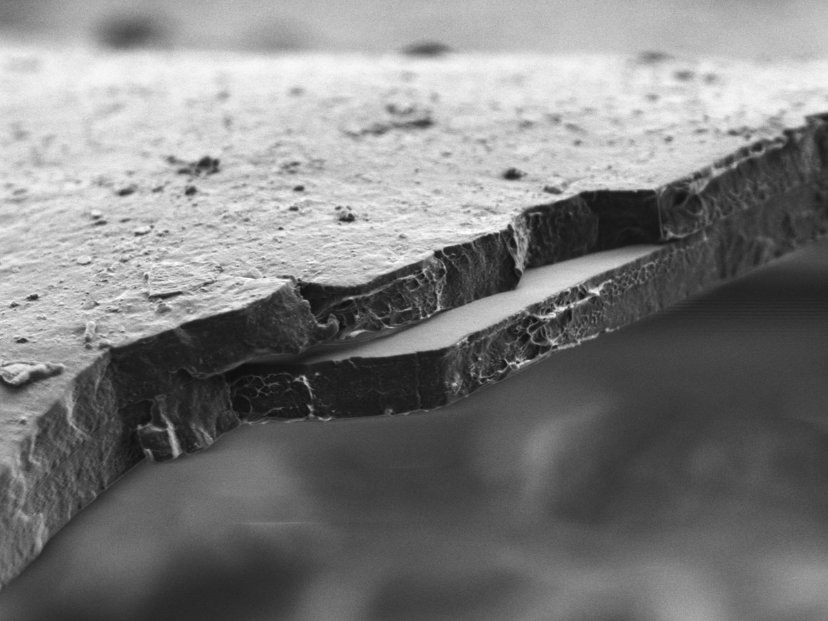
The use of ordinary Portland cement (OPC) as a binding agent in concrete contributes to 10% of global CO2 emissions. These emissions can be reduced, and concrete durability and mechanical properties can be improved by partially replacing OPC with supplementary cementitious materials (SCM). However, understanding the connection between the more complex microstructure of SCMs and their macroscopic properties is crucial. A new non-destructive testing method has been developed to investigate the mechanical properties of microscale-sized cement paste prisms. This involves using a specialized indentation technique to perform unconfined uniaxial compression at the microscale. This image of tested samples was produced using a scanning electron microscope, and it shows that one of the prisms collapsed into the shape of a heart.

Dangerous liaisons
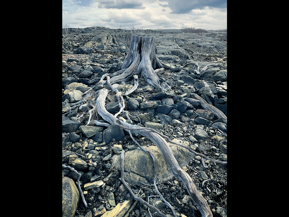
The zebra mussel is an invasive Eurasian mollusc that can harm North American-native mussels through intense biofouling—that is, by attaching to and overgrowing the North American mussel shells—which can cause suffocation, starvation, energy loss and subsequent death. My research examines the impacts of zebra mussel biofouling on native mussels in an invaded Quebec lake whose water chemistry was thought to be suboptimal for supporting a dense zebra mussel population. An exploratory scuba dive in the lake discovered an alarming level of biofouling of the same magnitude observed in a few sites deemed to be optimal habitats for zebra mussels. The zebra mussels have caused the collapse of native mussel populations, and my research results will inform and possibly revise risk assessments to predict which habitats’ native biodiversity is most susceptible to a zebra mussel invasion.

One blast’s fragile story
Gabriel Ciccarelli
A soot foil is a soot-covered metal sheet used to measure the size of detonation cells. The seemingly fragile gossamer featured in this image is a footprint left on a soot foil by a detonation (a scientific phenomenon of immense power and destruction). Transverse waves are formed due to the complicated chemical reactions that release heat and drive detonations, and as a result, leave this sophisticated pattern on the soot-coated surface. Understanding the mechanism underlying the emergence of such dark lines on the foil is a crucial part of our research, because unleashing the potential of detonations could lead to numerous advances, from harnessing their power for propulsion to enhancing our response to blast-wave disasters.
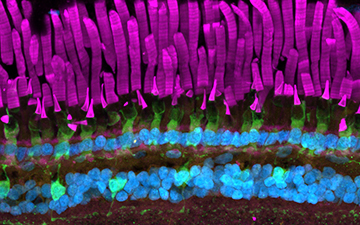
Frog retina
This image shows the retina of an African clawed frog (Xenopus laevis). The retina is a neural layer that lines the back of the eye and is responsible for detecting light and transforming it into visual signals. The retina is a highly organized tissue that is made up of five different classes of neurons and supporting glial cells. The rod and cone photoreceptor outer segments can be seen in magenta, and these are the parts of the photoreceptor neurons responsible for capturing light. The top green layer (directly below the magenta rods and cones) is made up of cone inner segments, and the green layer further below shows another retinal neuron type called bipolar cells. Muller glial cells and cytoskeletal components in neighbouring cells called the retinal pigment epithelium are labelled in orange at the very top of the image. Lastly, the blue spots indicate retinal neuron cell nuclei.

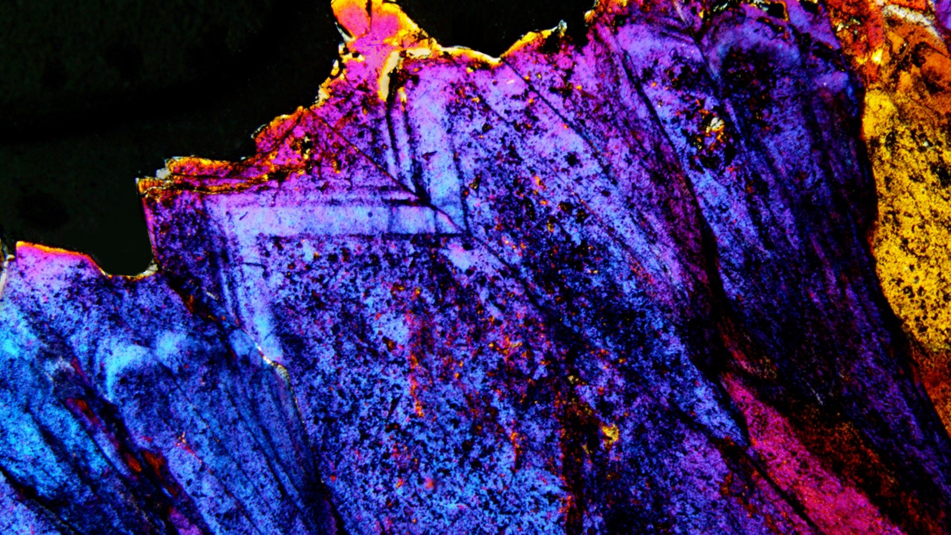
Geologic fluids in a crystalline time capsule
Guoxiang Chi
Fluid inclusion analysis is a means of looking millions of years into the past for insight into the compositional information of fluid responsible for forming mineral deposits. It can also be used to determine the pressure and temperature conditions under which critical metals were deposited. This image shows drusy quartz (crystal) within a sandstone sample from the Maw zone rare-earth element deposit in the Athabasca basin of Saskatchewan, containing preserved fluid that was once abundant in this region, but is now long gone. We can see stripes running along the crystal boundary, which are clusters of microscopic liquid and vapour droplets entrapped in bands as the crystal grew outwards in open spaces over thousands of years. The goal of our study is to use these fluids to provide insight into how these deposits formed and improve the parameters to be used in the hunt for new deposits.
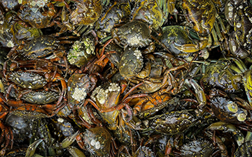
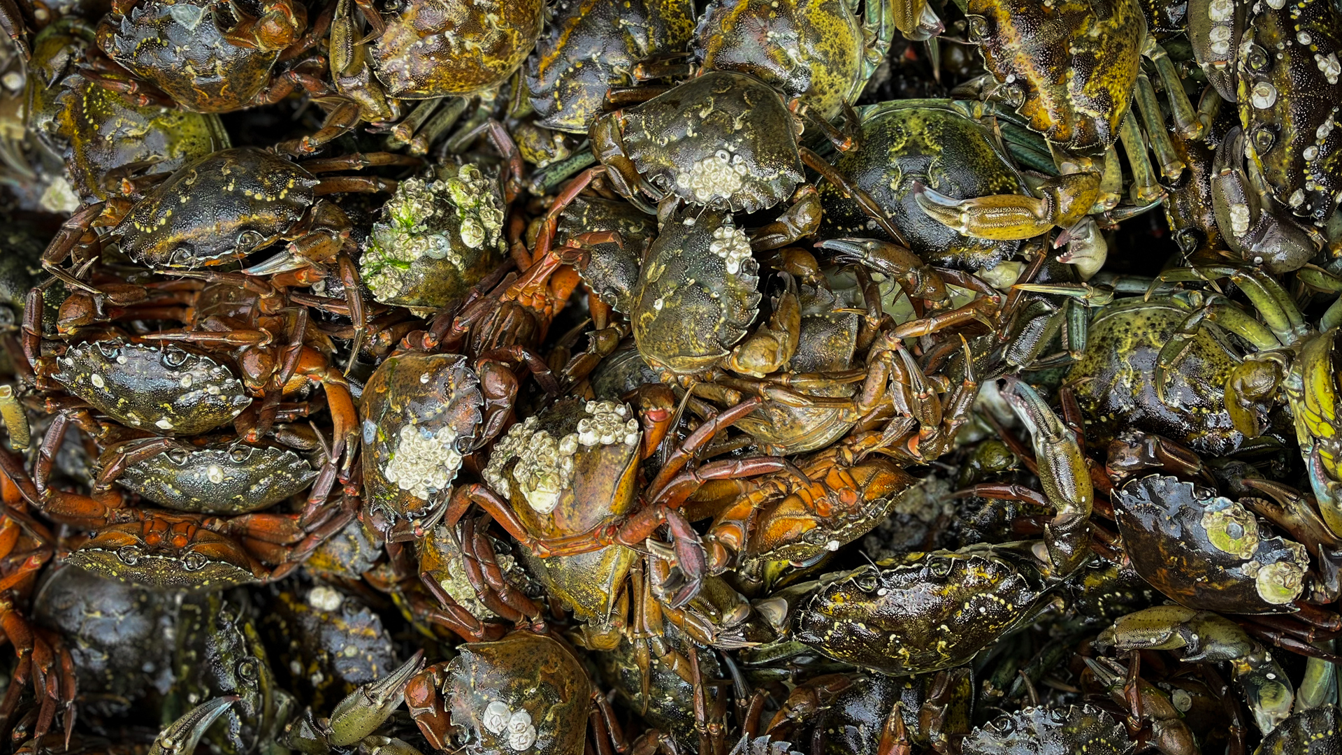
Green crab galore
The European green crab (Carcinus maenas) has been invading Canadian coastlines since the 1950s. With no sign of their invasions slowing down, it is imperative that we predict and understand their effects on ecosystems and the economy. Functional response experiments, which examine consumption by predators in relation to prey density, are a useful way to assess the potential strength of novel predator-prey relationships. These experiments often use only males, ignoring the potential influence of the invader’s sex. We investigated the potential differences in foraging between female and male European green crabs using functional response experiments in collaboration with the Coastal Restoration Society. We identified a potential overestimation of the impact of green crabs on their prey as a result of only focusing on male crabs. This could result in changes to management strategies for dealing with green crabs.

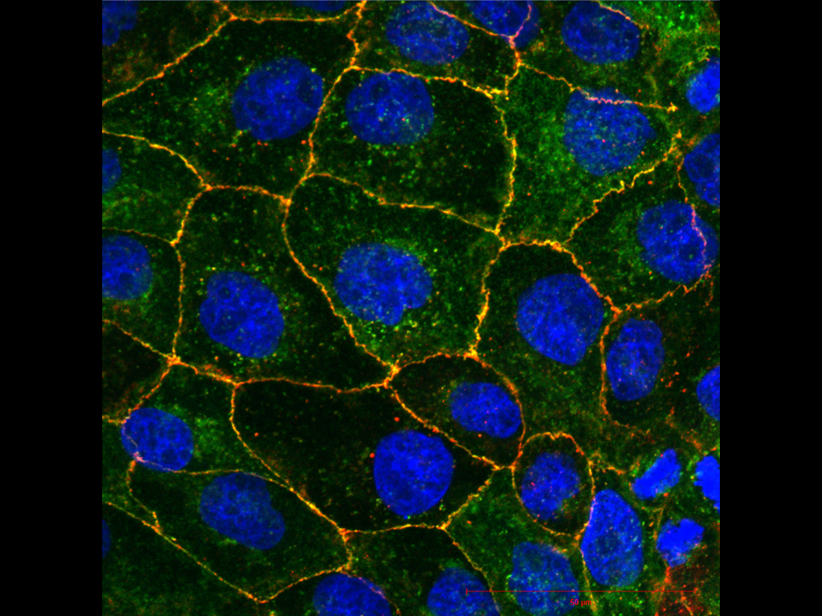
Kidney cells expressing CLDN8 protein
Tight junction proteins are important molecular components of cell-cell adhesion complexes that function as both ion pores and barriers between cells in different organs. Claudins are a large family of these proteins that allow ions like calcium to be selectively reabsorbed in the nephrons of the kidney. This image shows MDCK II cells (a dog’s kidney cell line) expressing human claudin-8 (CLDN8), which has been tagged with a green fluorescent protein. The cell nuclei are seen in blue, and the red indicates an antibody to the ZO-1 protein that has been used to mark the tight junctions of the cells. The yellow areas show the co-localization between the claudin and ZO-1 at the tight junctions, which is being used to compare with mutant versions of CLDN8 that may fail to localize to the tight junctions. This research is all being used as part of a larger study on claudin mutations found in a cohort of young patients with recurring calcium based kidney stones.

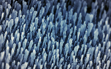
Lost in a microscopic bird’s nest
The bird’s-nest-looking material shown in this scanning electron microscopy image is a 100-nm-zinc oxide (ZnO) coating on a carbon paper substrate thanks to atomic layer deposition—a technique for growing highly conformal coatings that controls thickness at the monolayer level. Carbon paper is a commonly used anode material in lithium ion batteries, and its cycling capability and stability were improved with the ZnO coating. The author acknowledges Dr. Zoya Sadighi and Dr. Jeffrey S. Price for their contributions to this synthesis.

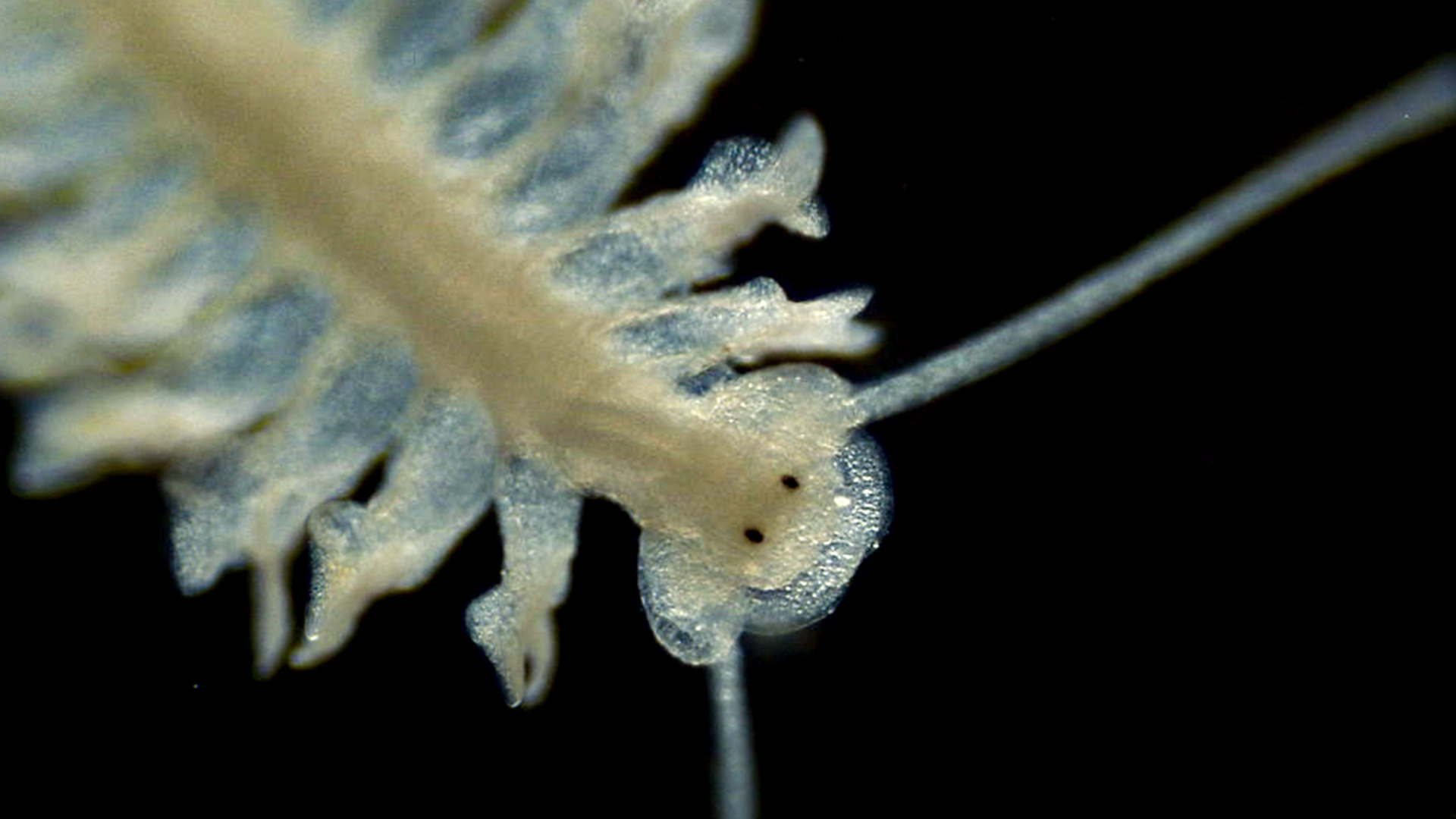
Ocean eyes
The ocean is full of microscopic creatures that we rarely think about, but they form the foundation of the marine food web that supports the largest marine animals! These microscopic creatures belong to a variety of phyla and are collectively known as zooplankton. The annelid worm in this image was swimming deep in the Bay of Fundy before being caught by our plankton net. At only a few centimetres long, it is nearly undetectable to the naked eye. Microscopy enables us to view it in enough detail to see its antennae, swimming appendages, and even its eyes. Analyzing zooplankton with a benchtop microscope helps us calibrate our underwater imaging devices, so that we can study zooplankton populations in finer detail across time and space. Using technology to gain a deeper understanding of zooplankton populations and how they affect marine megafauna can help inform effective marine conservation policies.

Only the sound remains
Shervin Foroughi
Muthukumaran Packirisamy
Cavitation can be defined as small gas bubbles being grown, oscillated and collapsed in a liquid medium while being affected by ultrasound waves. Traditionally, cavitation has been seen as a force of destruction in engineering, such as in the erosion of pump impellers, and it has evolved to be a fatal attack mechanism in nature. However, these bubbles can also be chemically active in certain sound wave and medium conditions in which the liquid medium can undergo physical transformation leaving a solid structure. Using a new 3D printing method called direct sound printing, we could tame cavitation and harness it for creation instead of destruction. This image shows the process in action: the sound waves are transmitted from the bottom to the printing medium. The bubbles are generated at the top, where the material can then be solidified.


