
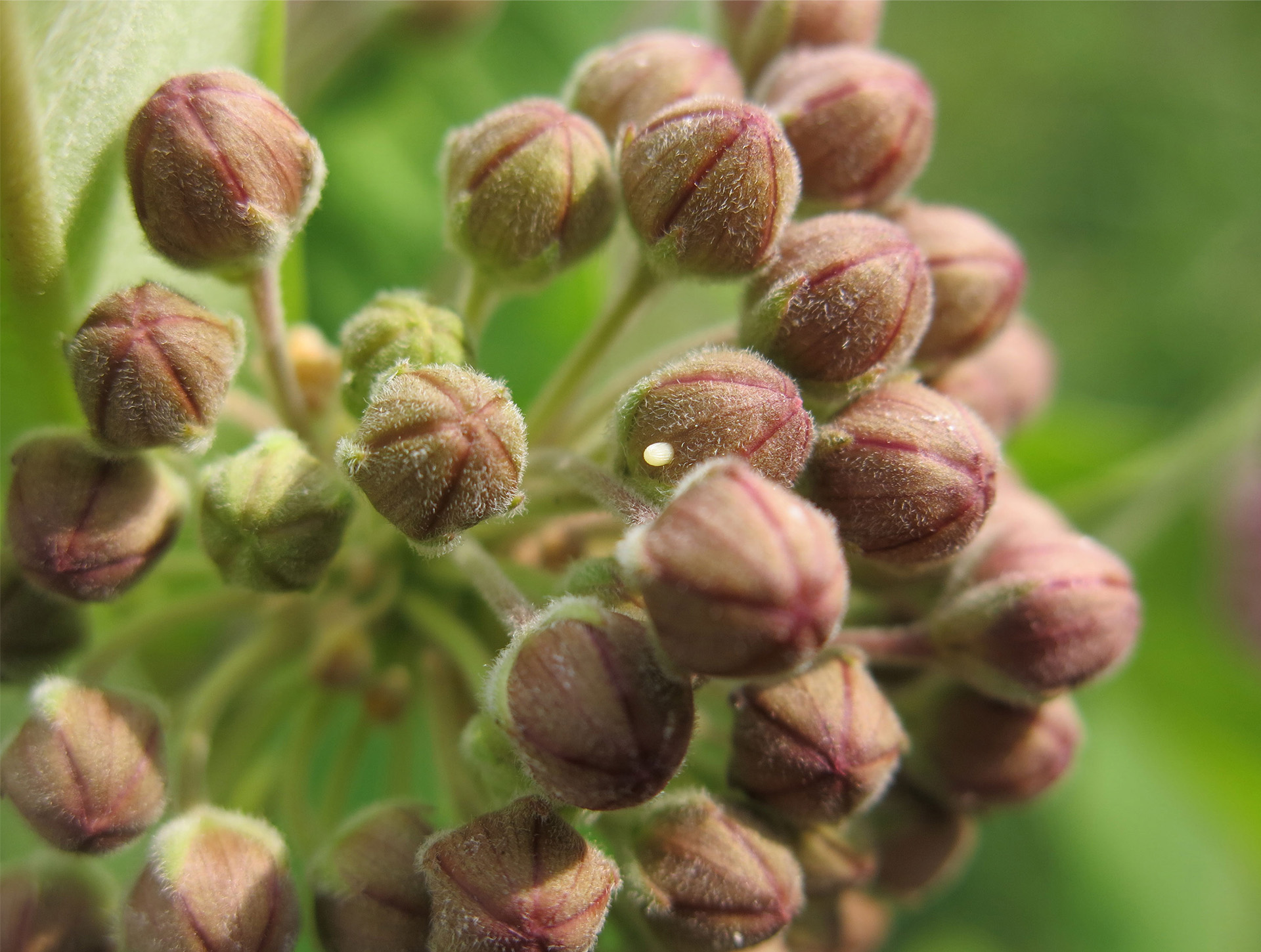
A snail’s first moments of sight
At only three days old, these tiny freshwater snail embryos have just begun seeing the world for the first time. While they don’t necessarily have true “eyes,” the eyespots (small black dots) and eyestalks of snails allow them to perceive light, pressure and chemical signals to orient themselves in their environments. Freshwater snails represent an important part of aquatic ecosystems by acting as nutrient recyclers. They are also excellent “canaries in the coal mine,” by helping us understand the impacts of the chemicals that find their way into our waterways and how we can better regulate them. Using time-lapsed macrophotography, we are studying how pollution can cause multi-generational effects long after the exposure period has ended.

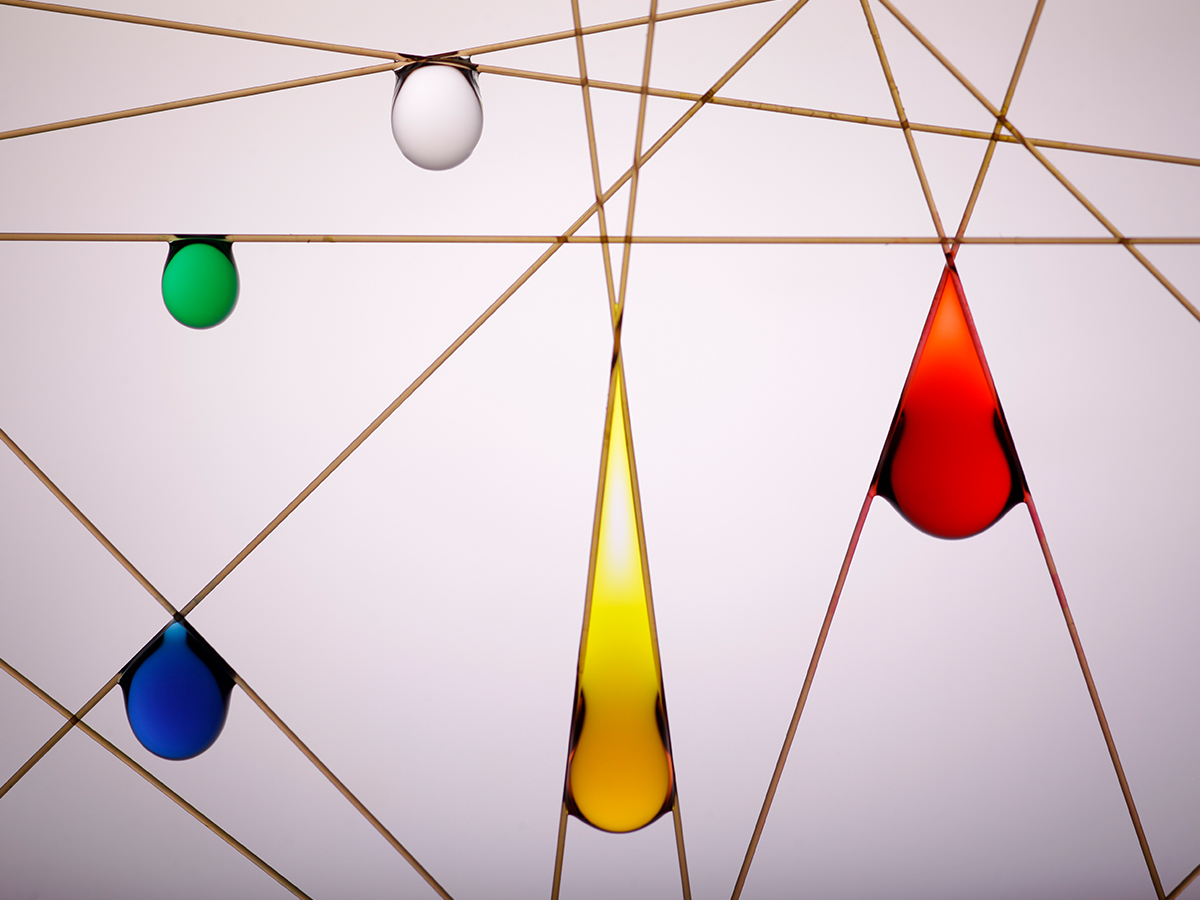
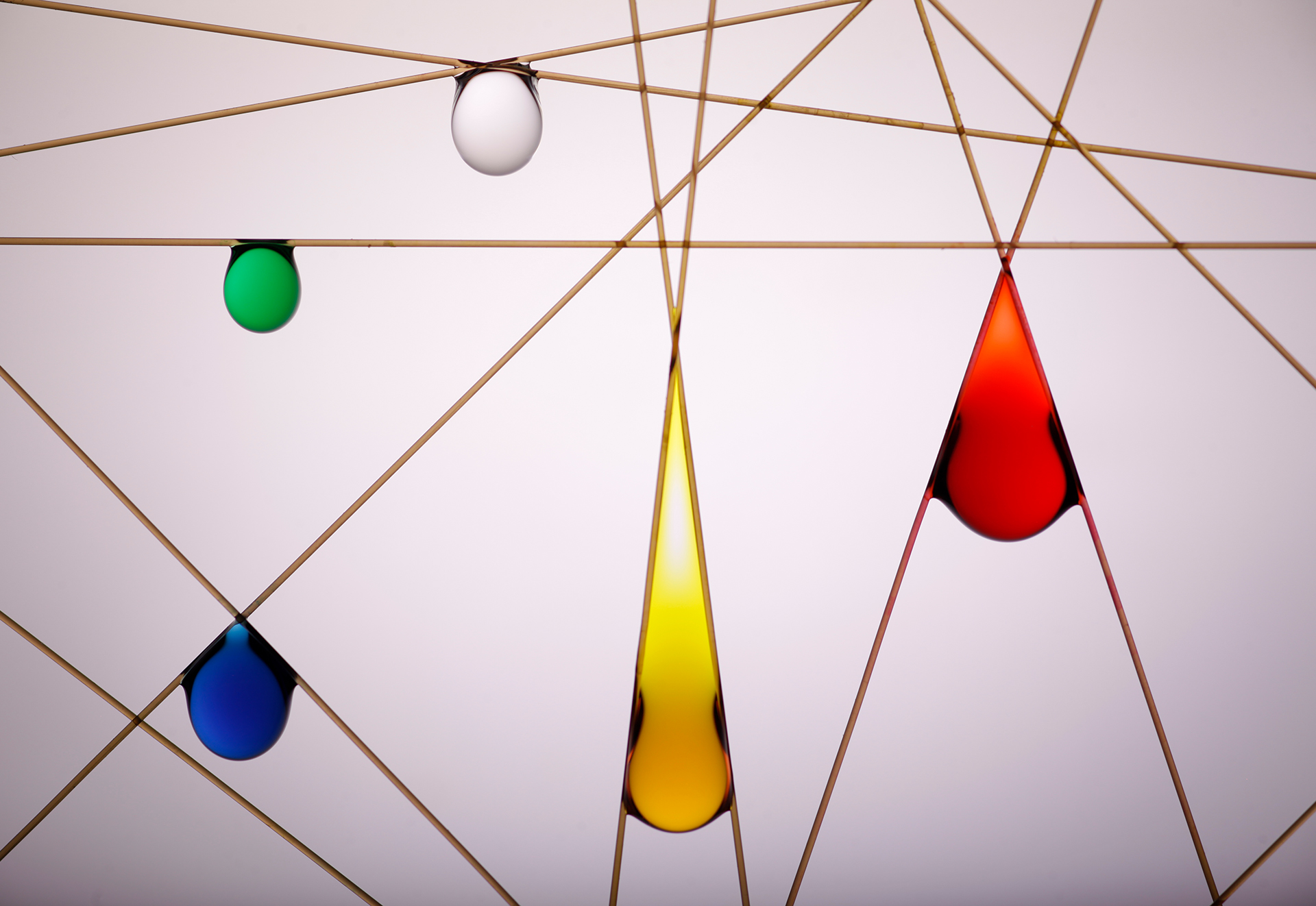
Composition with red, yellow, blue, green, and clear droplets
Long before photography existed, poets captured the mesmerizing behaviour of droplets: the ancient poet Du Fu penned “Heavy dew beads and trickles. Stars suddenly there, sparse, next aren’t,” and Jules Renard similarly observed “Quelques gouttes de rosée sur une toile d’araignée, et voilà une rivière de diamants.” Inspired by the huge droplets captured on the bifurcated leaf tips of cypress trees, we’ve found that the corner of two crossed fibres can hold significantly more water than a horizontally placed fibre. At 10 μL, the green droplet suspended on the horizontal fibre in this image holds the least water. As we decrease the angle between the fibres, you can see that the maximum droplet volume increases. The 89° angled fibres hold a blue droplet of 27 µL, and the 36° angled fibres maximize the volume of the red droplet to 65 µL. However, decreasing the fibre angle further to 13° reduces the volume of the yellow droplet to 37 µL.

Malignant brushstrokes
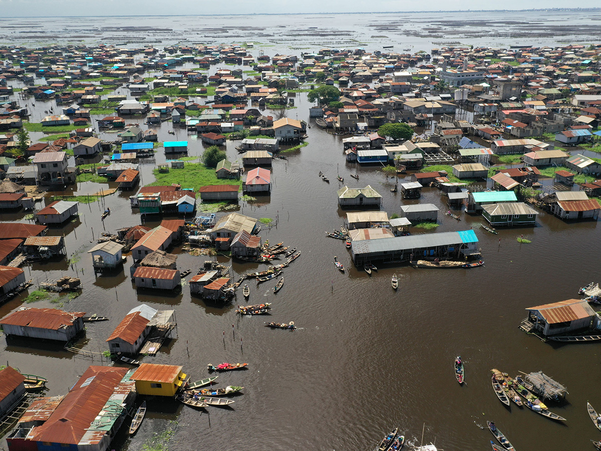
Human activity drives the intensity and frequency of blue-green algal blooms, which threaten aquatic biodiversity and the drinking water supply of millions. The transient and rapid emergence of these blooms into our lakes in late summer makes them difficult to monitor on short notice, particularly in smaller waterbodies. This drone image, taken from 100 m above the ground, shows my collaborators collecting water samples from an algal bloom in Dog Lake, a waterbody on the historic Rideau Canal system. The beautiful paint-like whorls seen from above hide a fetid and noxious “pea soup” that will eventually suffocate fish and other aquatic life when it decomposes in the fall. Using a combination of drone and environmental DNA monitoring, we are able to quickly assess the scale, movement and composition of a small bloom at the fraction of the price of satellite imaging or toxin assessment.

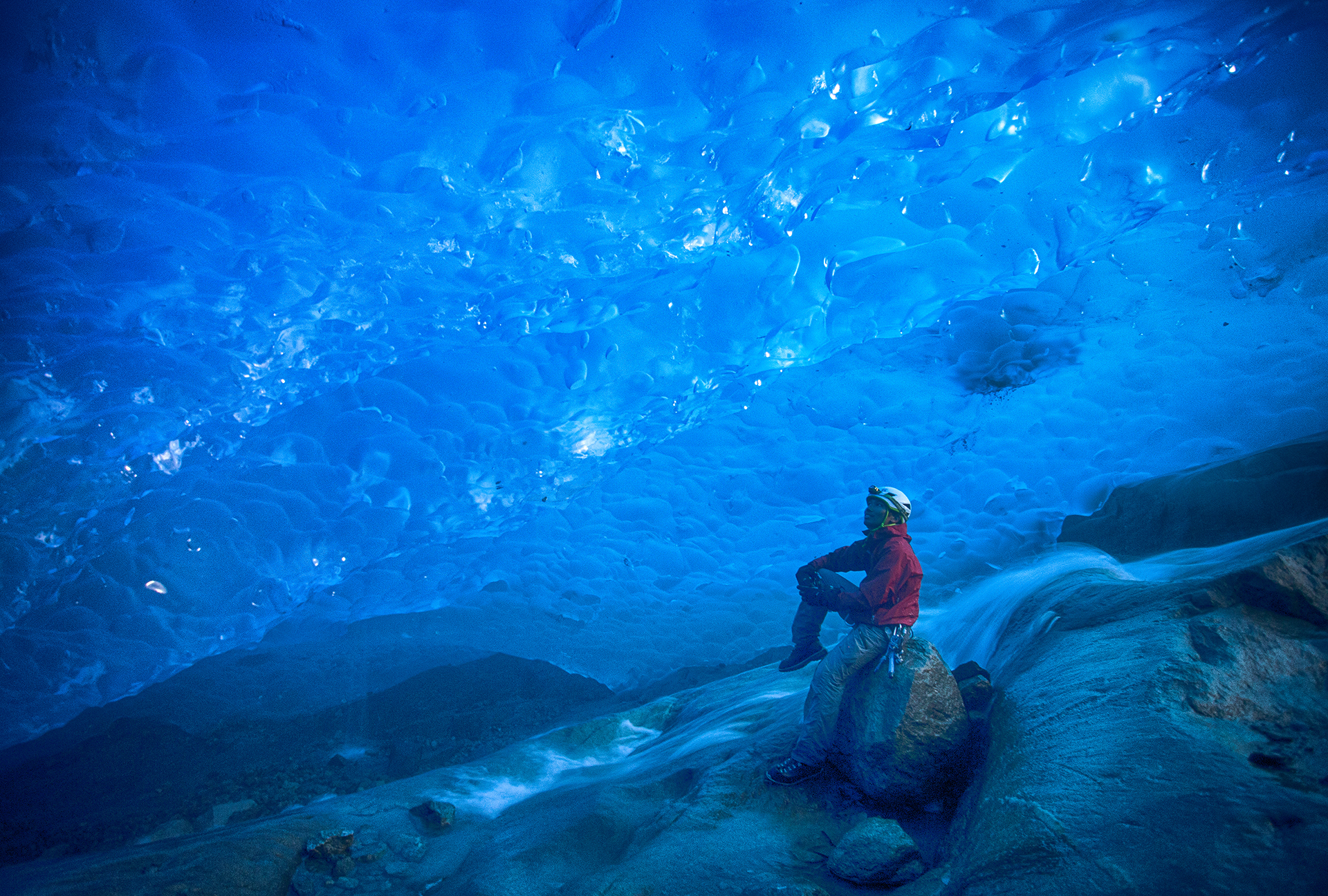
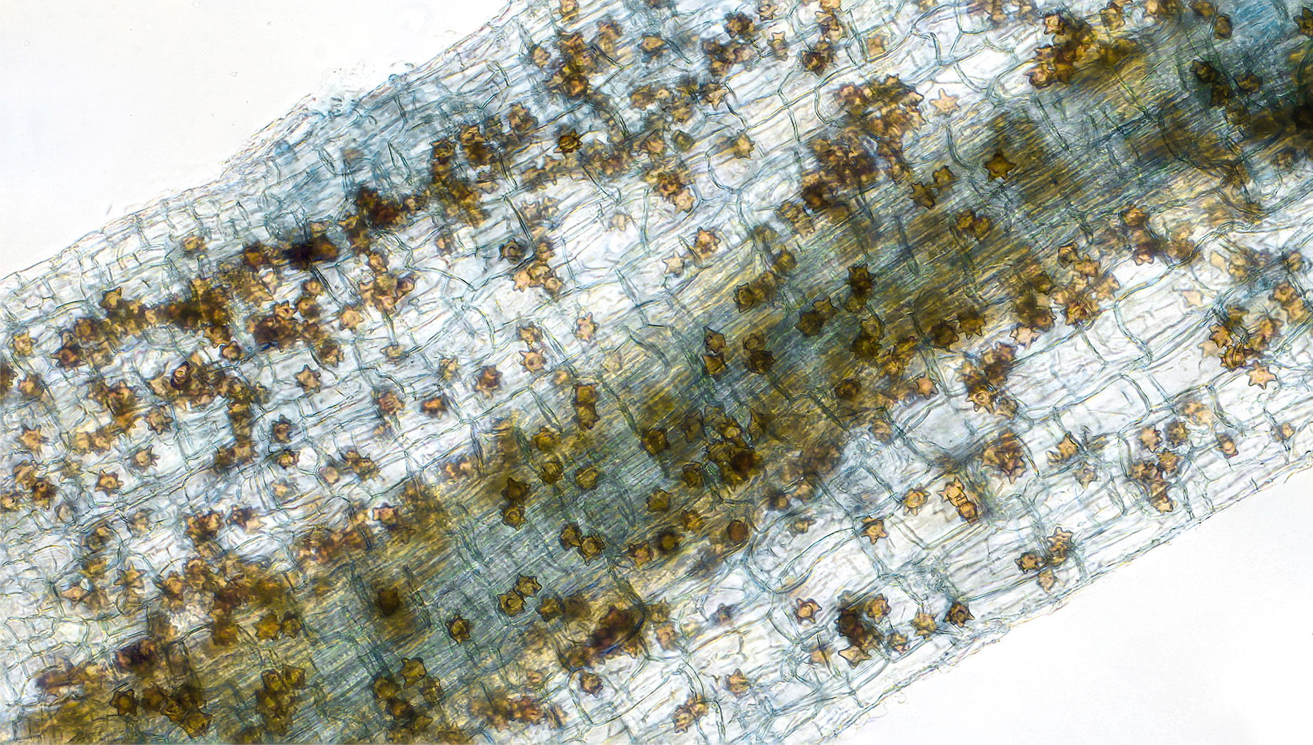
Mars tips its cap to the Arctic
Shown here are glaciers surrounded by false-color rock formations on Axel Heiberg Island in Nunavut. Note the yellow-orange areas, which are gossans. These surface deposits, rich in oxidized iron, indicate an acidic environment conducive to the appearance of certain lifeforms. You can also see a mineral whose crystalline structure can preserve traces of life: jarosite, a biomarker found on Mars! The researcher uses ground-based analyses to explore a method of identifying these caps on satellite images of the Canadian Arctic, knowledge that will be applied to satellite images of the Red Planet.

A task that goes unnoticed
"My work starts but it never stops." These are the words of a teacher correcting homework on a Saturday morning as her children play all around, forcing her to divide her attention. This image is part of a thesis on the relationship to work of preschool and elementary school teachers. The thesis looks at how, well before the arrival of telework brought about by the pandemic, a significant portion of tasks tend to spill over into other spheres of life. This work, which goes unrecognized, is hidden among the demands of daily life, making the border between personal and professional life permeable.

The rosette of the Thale cress
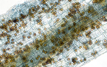
This microscopic plant assembly features the Thale cress, which grows naturally in this rosette form. Arabidopsis Thaliana is an ideal candidate for research in plant biology because of its rapid six-week life cycle. Here we see only cotyledons, the first leaves to emerge—except for an anther, where pollen grains are formed (top left). Each of the leaves is the result of specific research into its physiology, genetics, disease and weather resistance, reproduction or seed production, among other things.

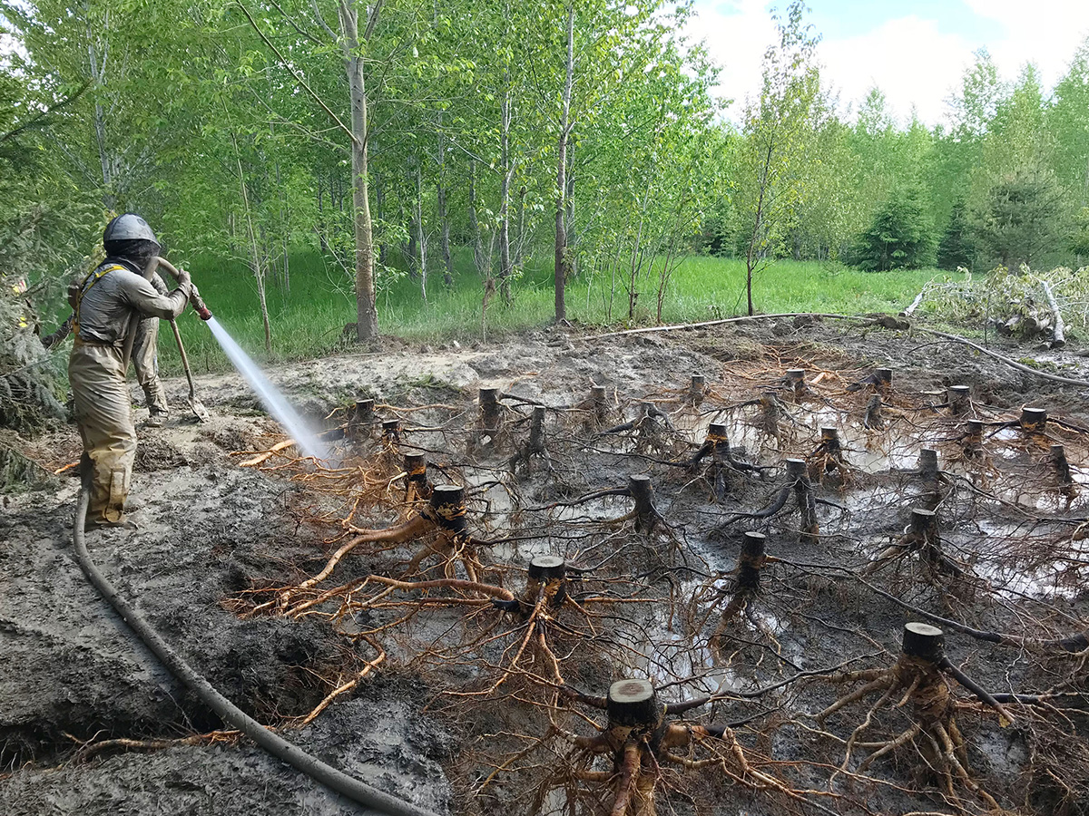
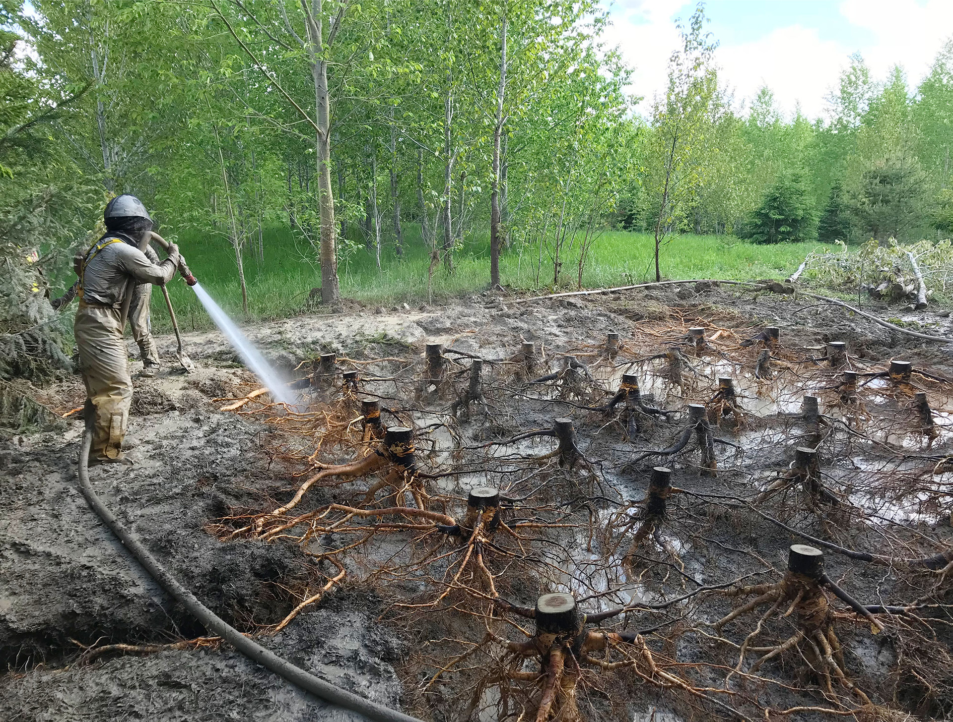
Hillside bog in green
The plants in this peat bog nestled in the Mont-Tremblant National Park is being inventoried to assess the impacts of climate change. To do this, botanists are identifying all the plant species in a 400 m2 plot (bounded by the triangle formed by the three squares), a tedious task requiring about three hours of work. But by using drone images and artificial intelligence analysis techniques, 20 minutes would be sufficient. The identification would be automated after training the model to recognize all the plants. Botanists would then supervise their "machine" colleague by validating the results.

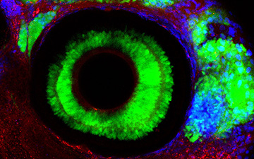
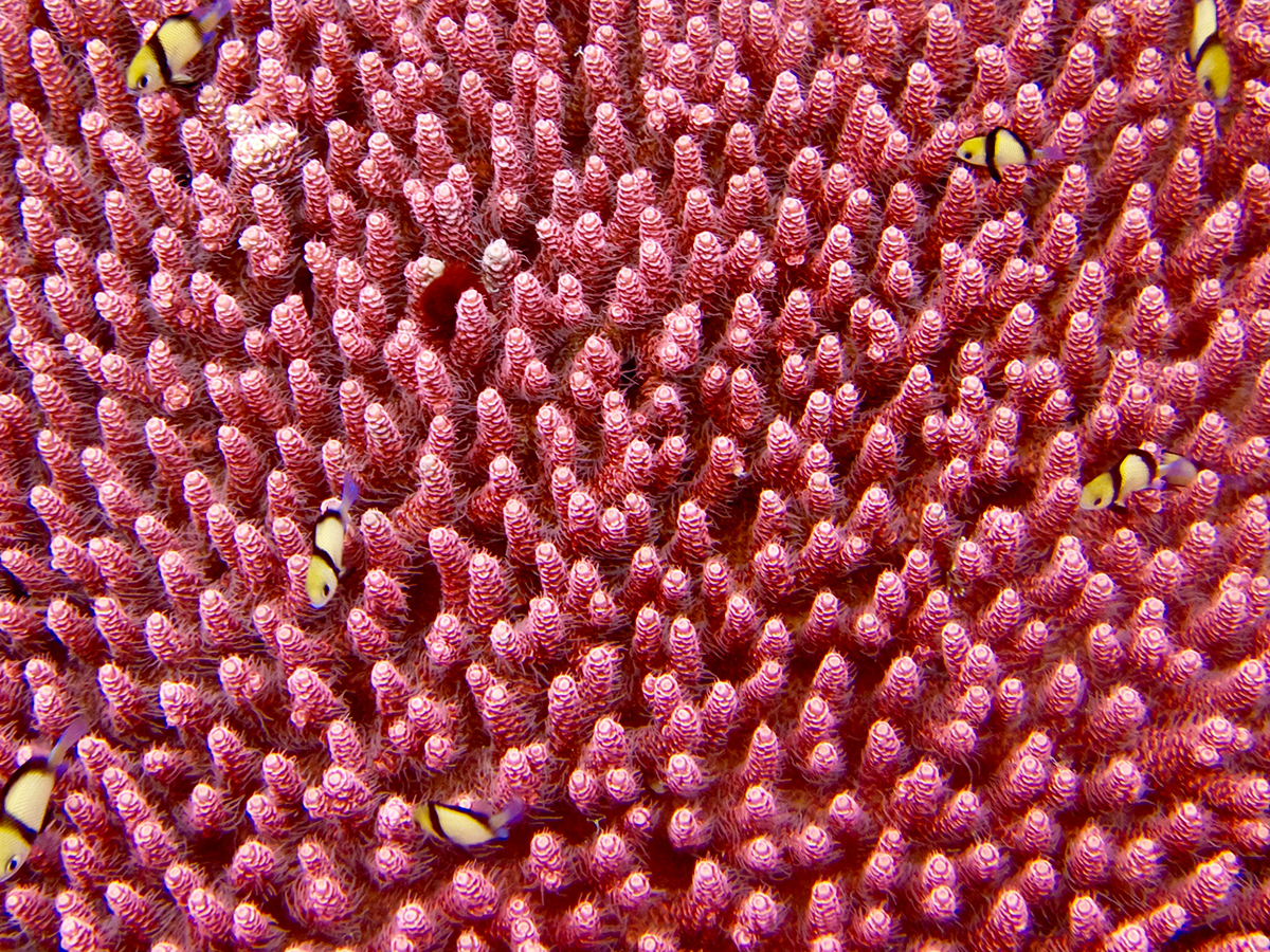
A smart, brainless jellyfish
Alexey V. Pshezhetsky
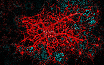
This fascinating maze of neurons comes from a region of the brain known as the hippocampus, which is essential for learning and memory. The neurons pictured here have been isolated from a transgenic mouse model of a pediatric neurodegenerative disease that causes cell death, leading to memory loss and dementia. These neurons were grown for 21 days on a cover slip in vitro and stained with a neuron-specific structural marker (white) to analyze neuronal phenotype MAP2. We are developing a series of treatments to restore the deficient protein that causes this disease. The affected neurons in this image were infected with a synthetic virus that overexpresses the deficient protein (red). This process leads to recovery and the establishment of healthy inter-neuronal connections.

Barley behind the scenes
Matthew Bakker
We can better equip ourselves to defend barley against potential fungi pathogens by improving our understanding of where it stores and synthesizes its essential survival tools. The aleurone layer—the honeycomb-like cells of this barley seed’s hull—serves as protection for the materials it needs for survival and germination. These tiny compartments house vital materials for the seed, such as enzymes, phenolic compounds and minerals, which are bound together in nutrient-rich globules (seen in red). Starch granules (the structures seen in yellow and purple) serve as important energy reserves for the growing barley plant. We used scanning electron microscopy to visualize the inner workings of this small seed and to locate the specific site of these important globules.

Bridging the gap
Energy storage solutions and especially lithium-ion (Li-ion) batteries play a central role in our pursuit for greener and increasingly efficient technologies. This image shows an electron micrograph of a modern Li-ion battery electrode. Exotic materials, such as carbon nanotubes (seen in blue) and carbon black (seen in red) have increasingly found their way into these electrodes. In the center of the image, you can see bundles of carbon nanotubes casting a web-like structure over the active material to create an electrically conductive network. These networks have been shown to improve energy density, charge rate, and life cycle of Li-ion batteries. Images like this help researchers develop a fundamental understanding of materials and electrode structure, which will lead to improvements in battery design.

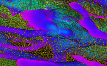
Cancer cells swimming in a microscopic paper forest
Much like humans, cancer cells are influenced by their surroundings. To develop new treatments against cancer, it is essential to understand how these cells are modified by neighbouring cells or even by their environment. My research consists of developing a model to answer these questions, so my team and I have created a biological “Swiss roll.” Instead of sponge cake and jam, we spread cells embedded in a gel (seen in cyan/red) onto a sheet of paper (paper fibers seen in green). Rolling the sheet of paper creates a 3D model with multiple layers of paper and cells, and by changing the type of gel, the type of cells, or even by mixing different cells together, we can recreate specific features of tumours. We can then use our model to study how cancer cells react to these changes. We hope our paper-based model will help turn the page on cancer research.

Do neural networks dream of falling snow?
This image shows 1.7 million vertices and 2.8 million edges, which comprise a snapshot in time of our deep learning neural network’s complex brain for calculating precipitation. As temperatures continue to rise, snowfall patterns are expected to change in complex ways with global impacts for springtime flooding, ecosystem development and water resource availability. High-accuracy precipitation models will allow us to better prepare for and mitigate against the consequences of a changing global climate. Each colour and cluster of vertices in this image represents a unique component of the neural network responsible for a specific task, and you can visualize how these pieces interact as neurons communicate across the network to predict rain and snow.
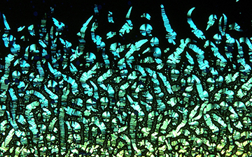
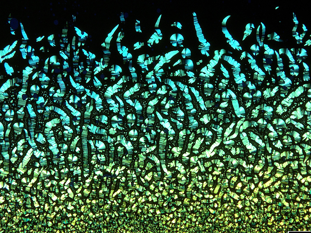
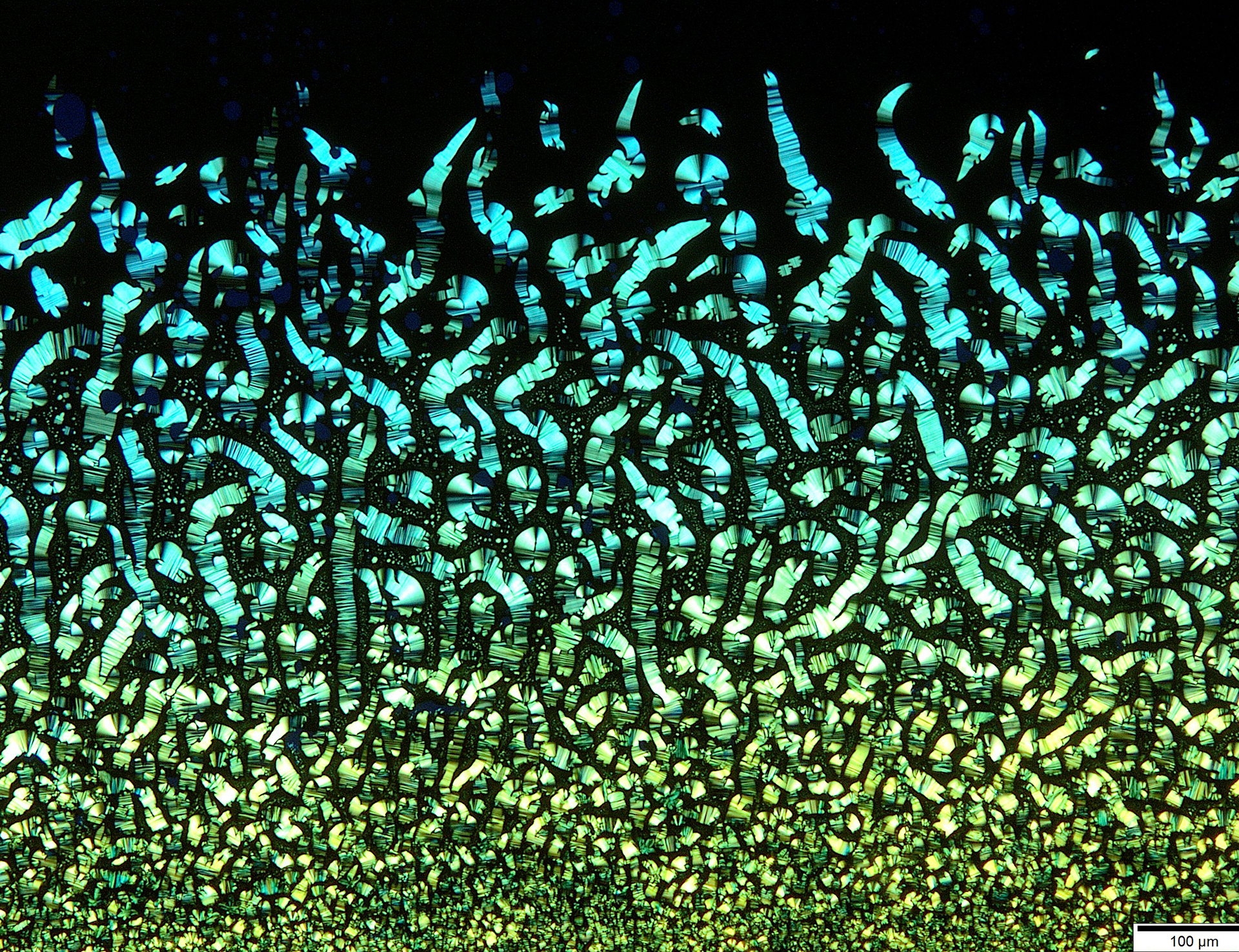
Microscopic emerald staircases
During my PhD I had the pleasure to work with liquid crystals, which are an intermediary phase between the states of solids and liquids. These liquid crystalline phases relay structural information and can be seen using polarized optical microscopy (POM), often with beautiful colours and textures. Pictured here is a novel compound I prepared, suspended in toluene and sandwiched between two glass slides. Using POM, we saw staircase-like features with vibrant green colours on the crystals.
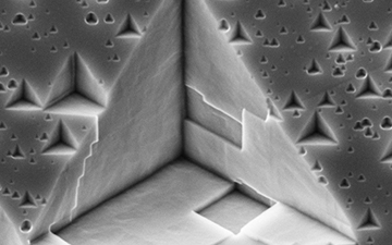
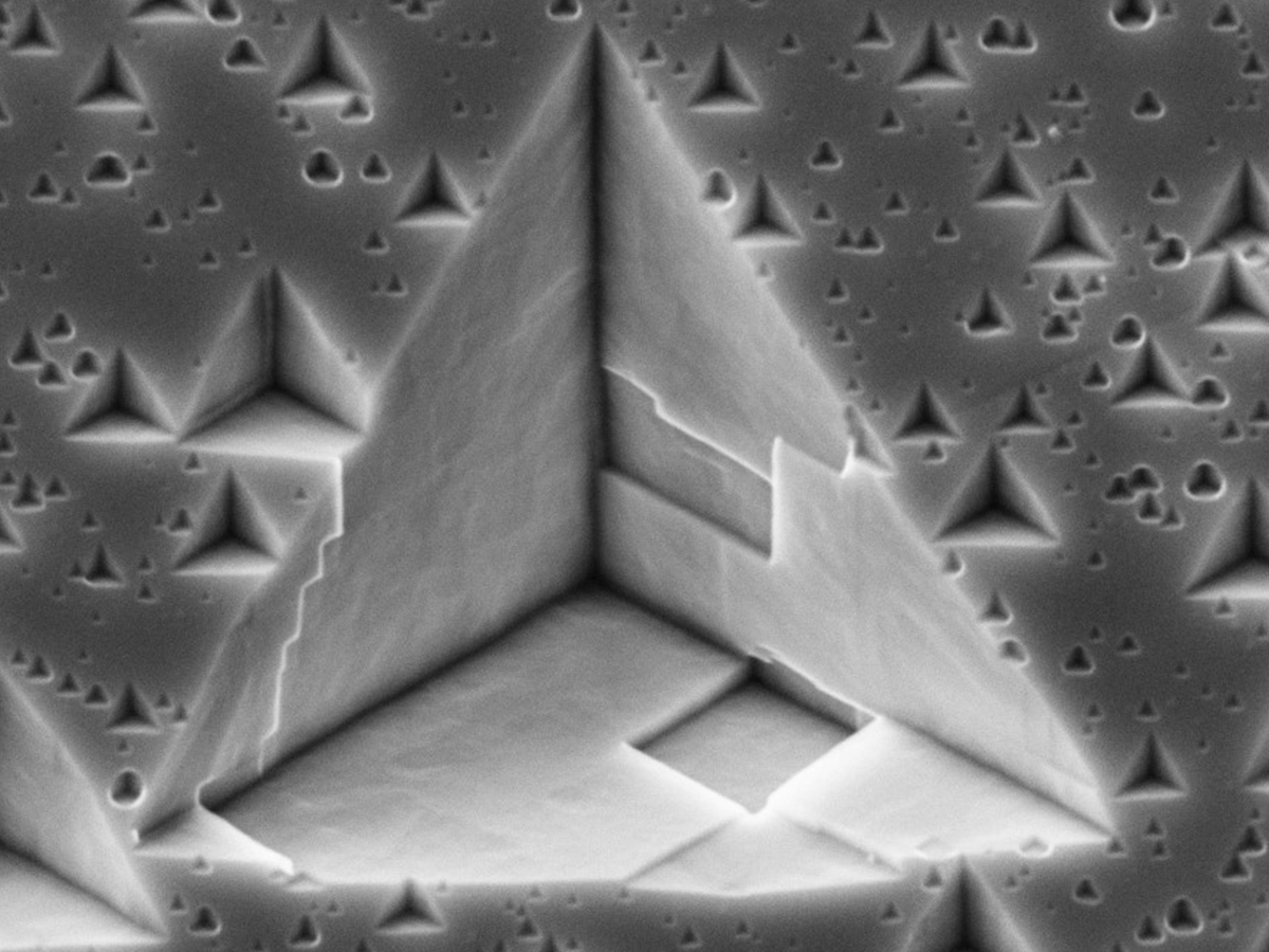
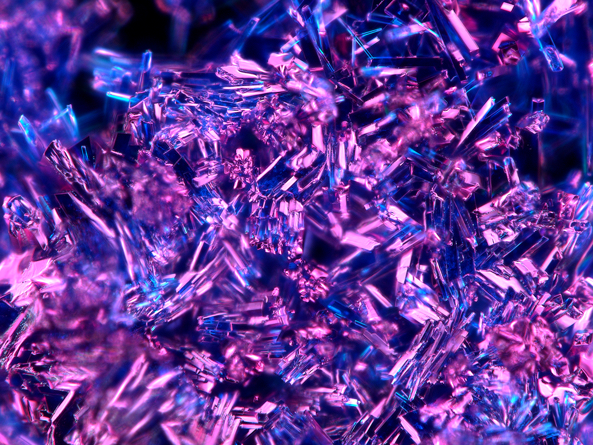
Resonance
When subjected to a chemical etch pitting solution, a cultivated single crystal aluminum material grown within a few degrees of its intended orientation uncovers dislocations. The shape and size of the exposed pits are relevant to the crystal’s orientation and dislocation nature. Since dislocations are an inevitable product of sample plastic deformation, any detected trail of pyramidal pits signals the incipient material’s mechanical loading history. The three-dimensional spatial arrangement of pyramids thus traces loading characteristics through plasticity (i.e., strain fields).

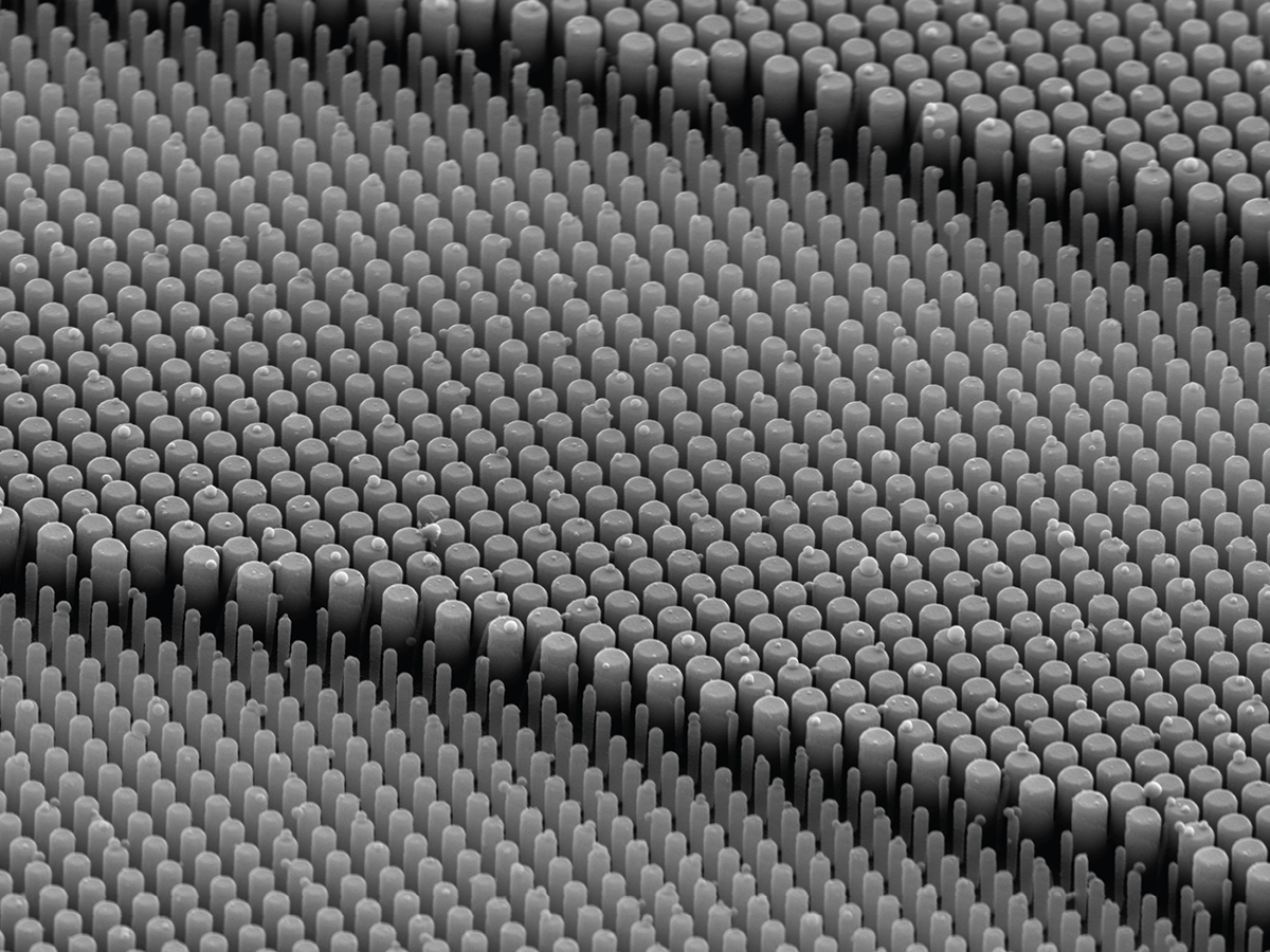
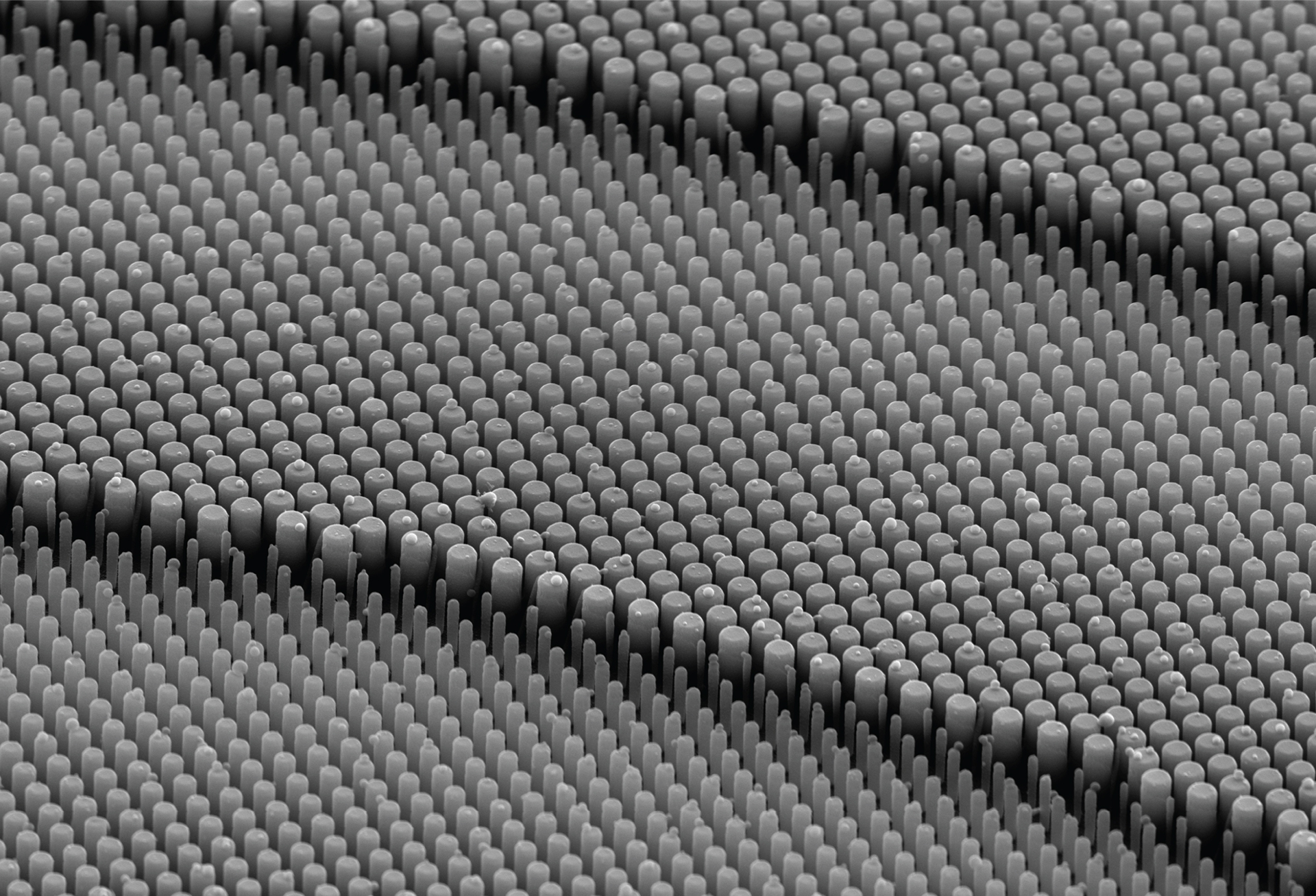
Fabricated nanostructures of a metalens
Metalens, an array of nanostructure optical elements, is a promising technology that could revolutionize optics by replacing conventional bulky lenses. By adjusting the shape, size and position of nanostructures, we can use metalens for complex imaging settings where conventional lenses fail to provide high-quality focusing. Our group, in collaboration with Harvard University, designed a metalens to incorporate into an endoscopic setting for live tissue imaging of internal organs. One-to-one comparisons of tissue images from both metalens and conventional lenses show metalens’ ability to capture images with noticeably higher resolution and more issue details. This research will ultimately enable a more sophisticated assessment of pathological changes, which could otherwise be easily overlooked by conventional lenses, at early stages of diseases like cancer.

Microfluidically generated salt crystal
Microfluidics is the study and manipulation of fluids at a microliter scale. Droplets can be manipulated using surfaces with different wetting characteristics. We generated magnesium sulfate salt crystals by evaporating a droplet of salt water on a microfluidically modified surface, and this image shows a perfectly circular salt crystal, 500 microns in diameter. While the image is coloured as a result of quality enhancements, salt crystals aren’t colourful.

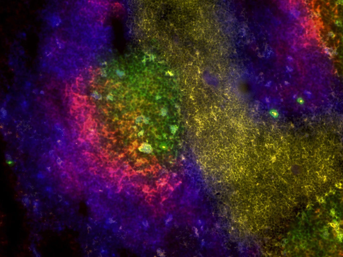
Mysterious festival of lights
This image shows highly abnormal neural stem cells of human origin. It is practically impossible to obtain neural stem cells from an intact human brain. However, recent progress in science has made it possible to convert normal body cells into pluripotent stem cells. These can then be differentiated into specialized stem cells (e.g., neural stem cells). The neural stem cells pictured here have a severe mutation that causes a rare and fatal neurodevelopmental disorder. Using organelle-specific fluorescent dyes, we can observe structures within the cells that present extreme abnormalities: DNA in the nuclei (blue), lysosomes (magenta) and mitochondria (yellow). Culturing and differentiating these mutant neural stem cells into neurons will help us understand the development of the disease and ways to treat it.
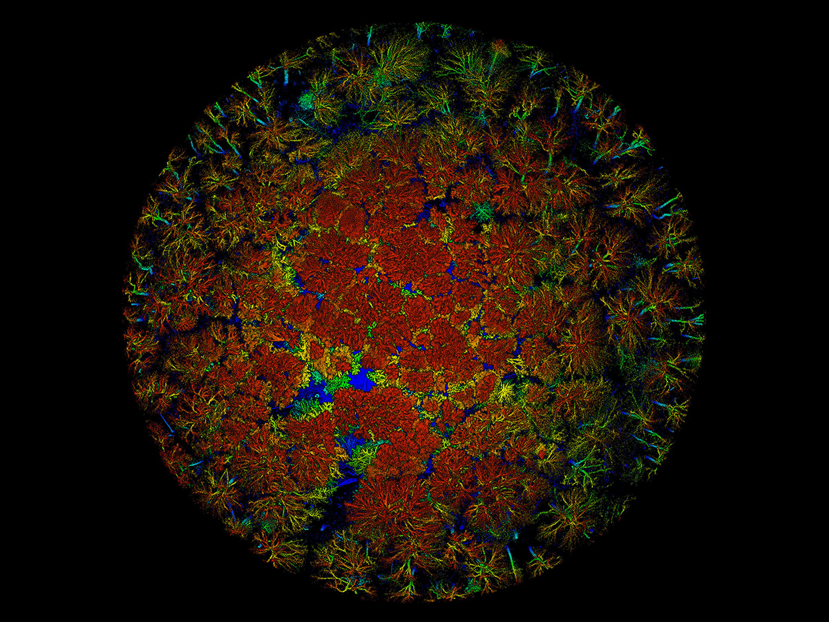
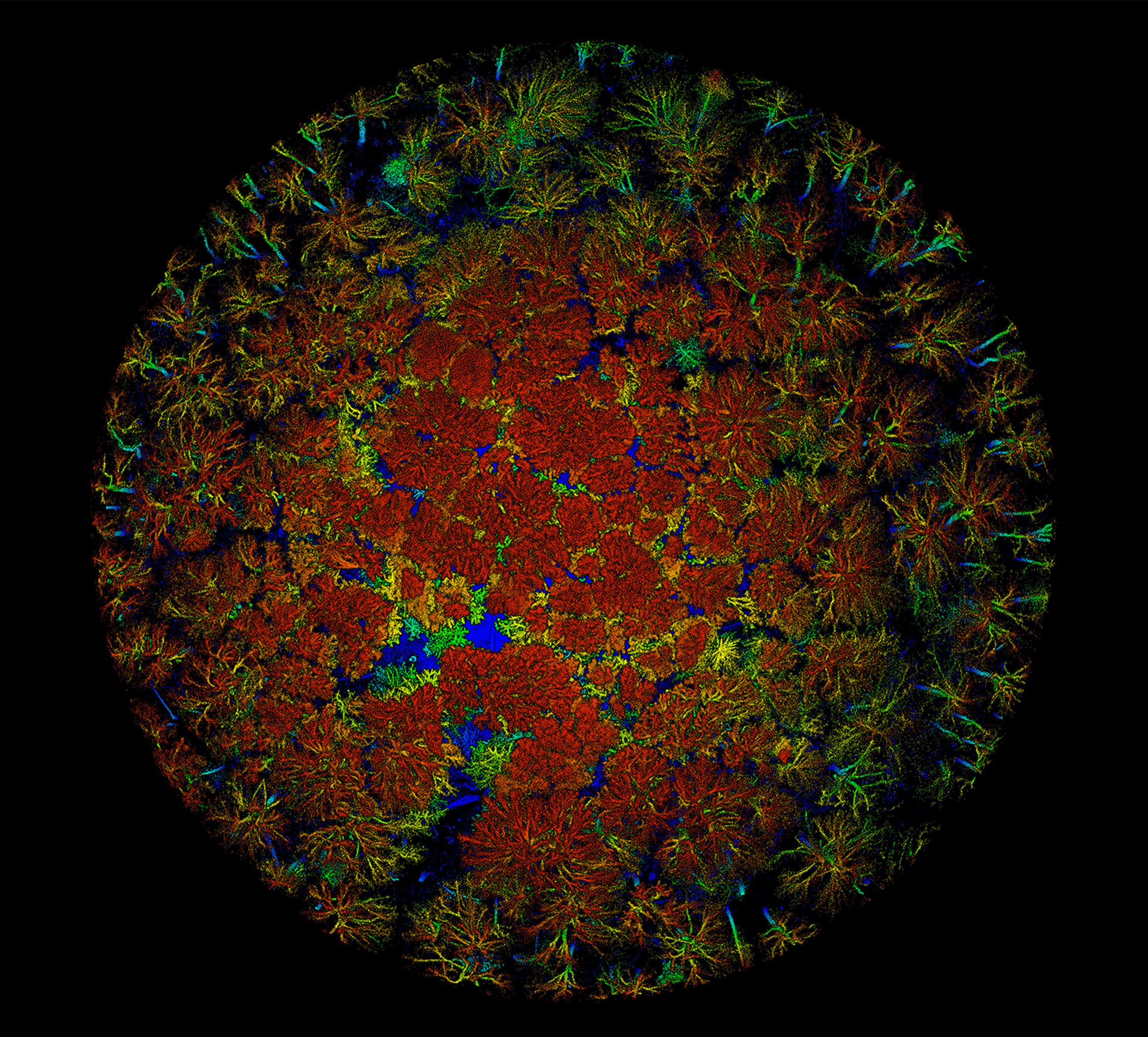
The very pores of osteoporosis
Janna M. Andronowski
Osteoporosis afflicts millions of people across Canada, and treatment and research around it costs billions—with an aging population poised to expand its impact further. New high-resolution approaches for the visualization and assessment of cortical bone porosity are emerging for both preclinical and clinical assessment of bone quality. Biomedical researchers at Memorial University are exploring changes in the vascular pore and cellular-level organization of human bone with age. This high-resolution 3D render of bone was generated using synchrotron radiation micro-computed tomography at the Canadian Light Source, Canada’s only synchrotron light source facility. This image shows the thickness of bone’s vascular pores from small (pink/blue; starting at 1.44 µm) to large (yellow/green; up to 464 µm), and the bone's cellular density (gold spots). Age-associated increases in porosity and decreases in cellular density may lead to increased bone fragility and decreased bone quality.
Playing tricks with lights to study myelin
Oligodendrocytes are specialized brain cells that wrap neurons with myelin (i.e., thin sheets of membrane that provide protection and insulation). When these cells are grown on a glass microscope slide, the myelin sheets flow from the cell’s appendages like oil over water and, like oil spilled on water, they interfere with the light passing through them. This image shows a live oligodendrocyte that has been modified to mark its body with a fluorescent protein. Between its intricate network of branches (red), we can see sheets of myelin (cyan) filled with striking light and dark whorls caused by light interference. Visualizing these cells as they grow helps us understand the complex cell biology that builds our brains and lets us test compounds to see if they boost myelin production, which will hopefully help to find new treatments for diseases like multiple sclerosis.

Crystallizing a new tomorrow
This microscopic image shows silicon phthalocyanine crystals as part of ongoing research into the thin-film formation of organic semiconductors for use in electronic devices. Organic electronic devices use carbon-based materials as the semiconductor, rather than traditional silicon or germanium. The moderate processing conditions of organic semiconductors enable low-cost energy efficient fabrication, unlike current technologies, which are costly and require energy-intensive manufacturing. Organic semiconductors also offer a myriad of innovative applications, including in medicine (such as artificial skin and point-of-need sensors), in bendable displays for televisions and smart phones, as well as in flexible transparent and portable solar cells for energy capture. My research focuses on the nucleation and crystallization of organic semiconductors to further the advancement of solution-based electronic manufacturing.


